
Glaucoma is a disease of the optic nerve, which transmits the images you see from the eye to the brain. The optic nerve is made up of many nerve fibers (like an electric cable with its numerous wires). Glaucoma damages nerve fibers, which can cause blind spots and vision loss.
 Glaucoma has to do with the pressure inside the eye, known as intraocular pressure (IOP). When the aqueous humor (a clear liquid that normally flows in and out of the eye) cannot drain properly, pressure builds up in the eye. The resulting increase in IOP can damage the optic nerve and lead to vision loss.
The most common form of glaucoma is primary open-angle glaucoma, in which the aqueous fluid is blocked from flowing back out of the eye at a normal rate through a tiny drainage system. Most people who develop primary open-angle glaucoma notice no symptoms until their vision is impaired.
Ocular hypertension is often a forerunner to actual open-angle glaucoma. When ocular pressure is above normal, the risk of developing glaucoma increases. Several risk factors will affect whether you will develop glaucoma, including the level of IOP, family history, and corneal thickness. If your risk is high, your ophthalmologist may recommend treatment to lower your IOP to prevent future damage.
In angle-closureglaucoma, the iris (the colored part of the eye) may drop over and completely close off the drainage angle, abruptly blocking the flow of aqueous fluid and leading to increased IOP or optic nerve damage. In acute angle-closure glaucoma there is a sudden increase in IOP due to the buildup of aqueous fluid. This condition is considered an emergency because optic nerve damage and vision loss can occur within hours of the problem. Symptoms can include nausea, vomiting, seeing halos around lights, and eye pain.
Even some people with “normal” IOP can experience vision loss from glaucoma. This condition is called normal-tension glaucoma. In this type of glaucoma, the optic nerve is damaged even though the IOP is considered normal. Normal-tension glaucoma is not well understood, but lowering IOP has been shown to slow progression of this form of glaucoma.
Childhood glaucoma, which starts in infancy, childhood, or adolescence, is rare. Like primary open-angle glaucoma, there are few, if any, symptoms in the early stage. Blindness can result if it is left untreated. Like most types of glaucoma, childhood glaucoma may run in families. Signs of this disease include:
- clouding of the cornea (the clear front part of the eye);
- tearing; and
- an enlarged eye.
Your ophthalmologist may tell you that you are at risk for glaucoma if you have one or more risk factors, including having an elevated IOP, a family history of glaucoma, certain optic nerve.
Conditions, are of a particular ethnic background (see section below), or are of advanced age. Regular examinations with your ophthalmologist are important if you are at risk for this condition.
The goal of glaucoma treatment is to lower your eye pressure to prevent or slow further vision loss. Your ophthalmologist will recommend treatment if the risk of vision loss is high. Treatment often consists of eyedrops but can include laser treatment or surgery to create a new drain in the eye. Glaucoma is a chronic disease that can be controlled but not cured. Ongoing monitoring (every three to six months) is needed to watch for changes. Ask your ophthalmologist if you have any questions about glaucoma or your treatment.
Glaucoma Evaluation
Because it has no noticeable symptoms, glaucoma is a difficult disease to detect without regular, complete eye exams.
During a glaucoma evaluation, your ophthalmologist will perform the following tests:
- Tonometry. Your ophthalmologist measures the pressure in your eyes (intraocular pressure, or IOP) using a technique called tonometry. Tonometry measures your IOP by determining how your cornea responds when an instrument (or sometimes a puff of air) presses on the surface of your eye. Eyedrops are usually used to numb the surface of your eye for this test.
- Gonioscopy. For this test, your ophthalmologist inspects your eye’s drainage angle—the area where fluid drains out of your eye. During gonioscopy, you sit in a chair facing the microscope used to look inside your eye. You will place your chin on a chin rest and your forehead against a support bar while looking straight ahead. The goniolens is placed lightly on the front of your eye, and a narrow beam of light is directed into your eye while your doctor looks through the slit lamp at the drainage angle. Drops will be used to numb the eye before the test.
- Ophthalmoscopy. With this test, your ophthalmologist can evaluate whether or not there is any optic nerve damage by looking at the back of the eye (called the fundus). There are two types of ophthalmoscopy: direct and indirect. With direct ophthalmoscopy, your ophthalmologist uses a small flashlight-like instrument with several lenses that magnifies up to about 15 times. This type of ophthalmoscopy is most commonly done during a routine physical examination. With indirect ophthalmoscopy, the ophthalmologist wears a headband with a light attached and uses a small handheld lens to look inside your eye. Indirect ophthalmoscopy allows a better view of the fundus, even if your natural lens is clouded by cataracts.
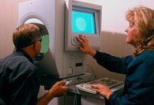
- Visual field test. The peripheral (side) vision of each eye is tested with visual field testing, or perimetry. For this test, you sit at a bowl-shaped instrument called a perimeter. While you stare at the center of the bowl, lights flash. Each time you see a flash, you press a button. A computer records your response to each flash. This test shows if you have any areas of vision loss. Loss of peripheral vision is often an early sign of glaucoma.
- Photography. Sometimes photographs or other computerized images are taken of the optic nerve to inspect the nerve more closely for damage from elevated pressure in the eye.
- Special imaging. Different scanners may be used to better determine the configuration of the optic nerve head or retinal nerve fiber layer.
Each of these evaluation tools is an important way to monitor your vision to help ensure that glaucoma does not rob you of your sight. Some of these tests will not be necessary for everyone. Your ophthalmologist will discuss which tests are best for you. Some tests may need to be repeated on a regular basis to monitor any changes in your vision caused by glaucoma.
Glaucoma: People of African and Hispanic Ancestry Are at Higher Risk
If you are of African or Hispanic ancestry and especially if you have a known family member with glaucoma, you are at a higher risk for vision loss from this eye disease.
Primary open-angle glaucoma is the leading cause of blindness among people of African ancestry, occurring at a rate four times higher than among Caucasian patients. It also occurs about 10 years earlier among people of African ancestry than among Caucasians and develops more rapidly. Studies show that in the United States, African Americans between the ages of 45 and 64 are approximately 15 times more likely to go blind from glaucoma than Caucasians with glaucoma in the same age group. Primary open-angle glaucoma is also the leading cause of blindness among people of Hispanic (and especially Mexican) ancestry, occurring at a rate approaching that of people of African ancestry.
It is not clear why people of African ancestry have higher rates of glaucoma and subsequent blindness than Caucasians. One factor may be that they are more susceptible to developing elevated IOP earlier in life, which is thought to contribute to optic nerve damage and eventual vision loss. Another reason may be that they are less likely than Caucasians to have early eye examinations that might detect and treat glaucoma. This also may be a factor in the increased rate of glaucoma among Hispanics.
Glaucoma causes no symptoms early in its course; you will not experience pain or vision changes while it is developing. The best way to protect yourself and your family members against vision loss from glaucoma is by being aware of your higher risk of developing this disease and by having regular eye examinations for glaucoma at appropriate intervals.
Recommended intervals for a comprehensive eye evaluation in people of African ancestry are as follows:
- 20 to 29 years of age: every 3 to 5 years;
- 30 to 64 years of age: every 2 to 4 years;
- 65 years and older: every 1 to 2 years.
It is also recommended that people of Hispanic ancestry have regular, comprehensive eye evaluations. This is especially important after age 60.
If you are diagnosed with glaucoma, please make sure to tell your family members and urge them to have an eye exam for glaucoma.